Anogenital Examination
All skin surfaces of the victim should be examined and the lesions that may occur during the attack should be determined. In cases where genital lesions are absent or ambiguous, lesions detected in the body can be important evidence of sexual assault. Before any examination of the lesions, samples should be taken with swabs and smear should be done and biological substances such as blood, saliva, sperm, etc. belonging to the aggressor should be searched. With physical examination, the location, nature, and extent of traumatic changes on the body should be specified and the lesions should be photographed in scale and color scale. It will be very helpful to do all these operations with a single mobile device during the examination. A multispectral device is needed to prevent the loss of evidence and to scan all biological materials that can be detected.
Examination of the external genital organs begins with inspection. The vulva, pubis, lateral parts of the abdomen, calf, and perineum should be investigated. It is recommended to examine this area with a device with optical zoom in terms of materials such as semen, blood, and hair. Hymen examination may be the only area with traumatic findings, especially in young girls and children. For this reason, this examination must be done completely and at once.
About the hymen, a small membranous tissue with no known biological function typically occupies a portion of the external vaginal opening in females. The appearance of the hymen changes with age and in response to hormonal influences. The prepubertal hymen is characterized as thin, translucent, and sensitive to touch. It becomes thickened, elastic, redundant, and accommodating in puberty, as the result of a physiologic increase in estrogen exposure. To evaluate these features, the structures that make up the vaginal canal should be photographed from different angles. In situations such as small children or mental retardation where the examination is difficult, it is very difficult to obtain clear images from different angles with a fixed camera. In such cases, the examination process can be performed much more easily with a mobile device such as a “Smartphone” capable of optical zoom.
Hymen examination should be done in a well-lit environment. In hymen examination, the shape, character, opening, characteristics of the edges, resistance and elasticity of the hymen, whether there is a laceration in the hymen, whether it is old or new, how many lacerations are, whether it extends to the vaginal wall and whether there are other traumatic findings are examined. When evaluating Hymen, it is necessary to distinguish between partial lacerations and congenital natural notches, and the distinction between old and new lacerations, and it is necessary to visualize the hymen tissue closely to make this distinction. The “Smartphone” can show the hymen tissue in great detail thanks to its high-resolution camera with optical zoom.
Injuries in the anogenital area can sometimes be very small. It is very difficult to detect microtraumas and to evaluate the healing processes of existing lesions with the naked eye. It is possible to detect invisible lesions such as ecchymoses in millimetric dimensions with optical zoom imaging devices. In addition, with a close examination, the healing stages and wound age can be determined in detail.
Imaging with a multispectral device is required to evaluate all these examinations well. For example, to see whether a detected laceration reaches the vaginal wall or not, it should be viewed from different angles and photographed. At this point, it can be challenging to perform this inspection with a fixed camera. This can be done easily with a mobile device with a good light source. In addition, the fact that this area is wet during the examination may lead to reflection and incorrect evaluations of the images taken. In this case, artifacts in the image taken with different filters should be prevented. A device that includes light sources of different colors and wavelengths and multiple filters, such as a “Smartphone”, meets the needs of this examination.
During the examination, all of the anogenital areas should be examined in detail. For patients that cannot cooperate with the examination, it is better to use a mobile device instead of positioning the person. “Smartphone” is a mobile device and it provides more free examination opportunities as it is not dependent on the outside of the cable, etc. In addition, the “Smartphone” has a built-in lithium battery and it provides hours of examination without the need for an external power source.
Images should be recorded while performing the anogenital evaluation. “Smartphone” can save images in high resolution as RAW format. High-resolution images provide the opportunity to examine again and more in detail either on the “Smartphone” or on the computer after the examination is completed. A RAW file is a collection of unprocessed data. This means the file has not been altered, compressed, or manipulated in any way by the computer. Therefore, the RAW format is the most reliable file format known in international justice systems and is accepted as evidence in courts.
Fig 1: Acute complete laceration with ecchymosis at 7 o’clock extends to the vaginal base and Acute partial lacerations with ecchymosis at 4 and 5 o’clock
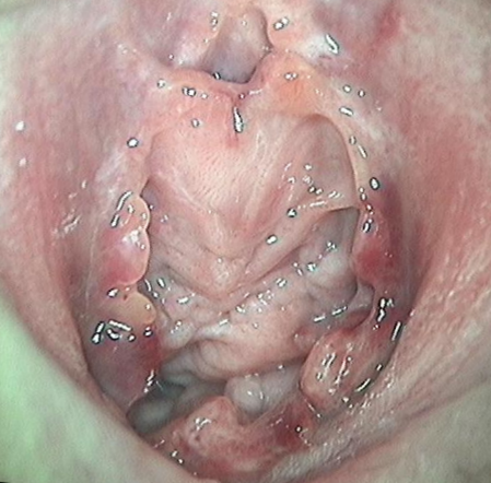
Fig 2: Acute partial lacerations with ecchymosis at 4 and 9 o’clock
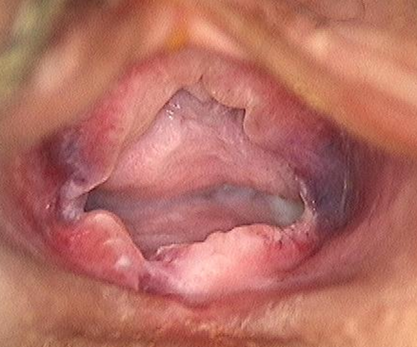
Fig3: Differentiation of old complete and partial lacerations and their extends to the vaginal base
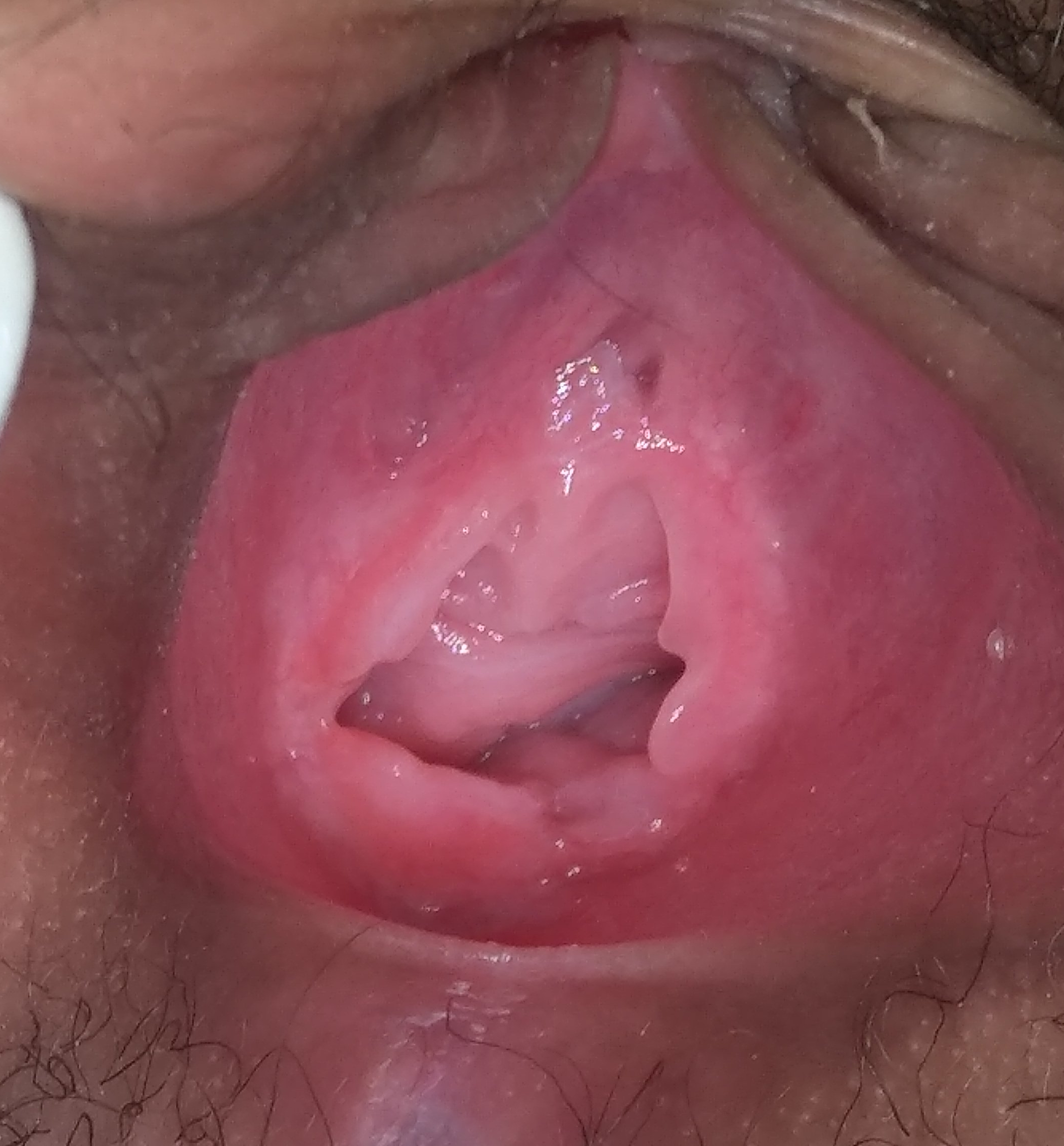
Fig4: Suspected lesion at 7 o’clock
High magnification with optical zoom shows a continuing line for hymenal rim and residual hymenal tissue at the 7 o’clock location
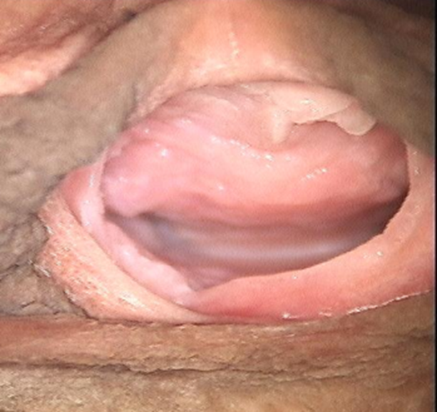
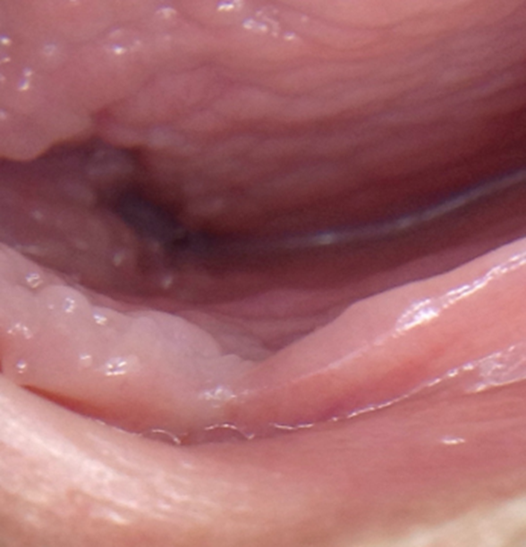
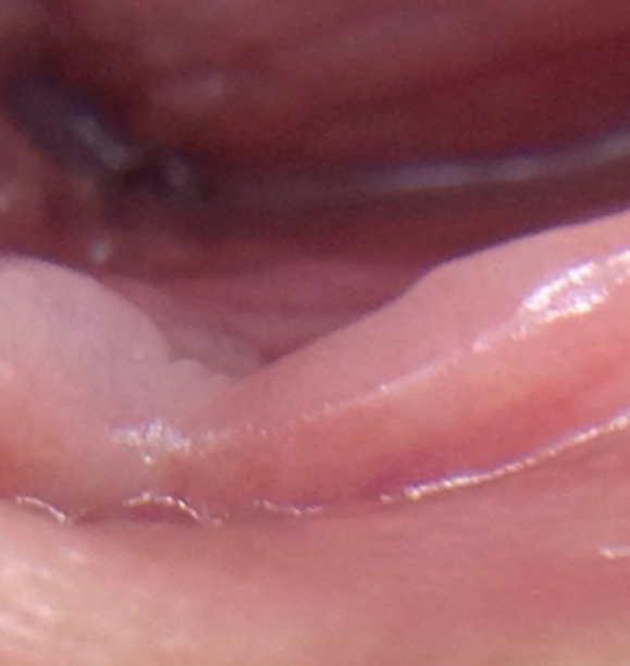
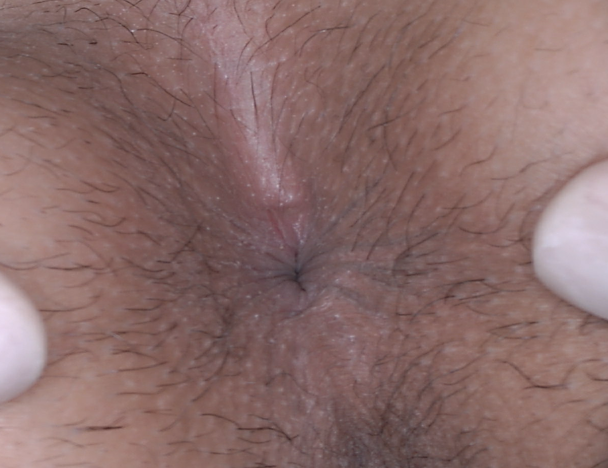
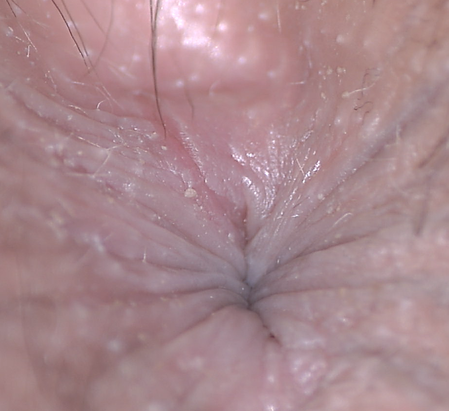
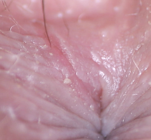
Fig 5: Samples of live magnification of anal fissures with optical zoom