In recent years, a growing number of technologies have been integrated into investigations involving the examination of bodies at crime scenes and in autopsy rooms. These technologies do not replace the work of the forensic pathologist, whose role is highly specialized and essential in providing primary information about the condition of the body and the changes within it. However, technologies assist in making the work more objective, easier, and of higher quality. These technologies can be associated with the automation of processes, documentation management, visualization, analysis and measurements, preservation of evidence, and more. Visual technologies primarily assist in two key areas:
- Improved and Objective Documentation: These technologies facilitate better documentation of findings, their archiving, and accessibility for later quantitative and qualitative analysis, including measurements and interpretation.
- Detection of Non-visible Evidence: They aid in identifying changes and traces that are not visible to the naked eye or are located in difficult-to-access areas, with the goal of finding evidence, understanding the mechanisms involved, and reconstructing events.
In any investigation, it is crucial to find all traces that can serve as evidence, whether they have positive or negative value. These traces can confirm or refute a particular theory, prove guilt or innocence, assist in identifying individuals, locations, and objects, including weapons. The absence of traces is also valuable, primarily in describing mechanisms, and in determining the presence or absence of guilt in a suspect.
In forensic science, the principle of Locard holds significant historical and practical importance: every contact leaves a trace. Any person present at or passing through a crime scene leaves traces, or the scene leaves traces on them. These traces, once analyzed, can become evidence and solve a case. Often, a microscopic trace may be the only link between the perpetrator and the victim, or between a specific weapon and the injuries it caused. Therefore, it is of utmost importance to detect even the smallest and sometimes invisible traces. On the other hand, we must also ensure that, after thorough examination, no traces are left on the subject under investigation (which could be the crime scene, body, or other objects). It would be dangerous for justice if, after an investigation, we declare to the investigative authorities and the court that no traces were found, only for them to be discovered later or perhaps never at all. An innocent person could be imprisoned, or the victim may never receive justice.
Technologies
One of the most popular and important technologies introduced in forensic autopsy practice is the so-called virtual autopsy, or postmortem imaging, using computed tomography (CT) or magnetic resonance imaging (MRI). These medical technologies are used to examine the body and all its internal structures in layers, to identify pathological and traumatic changes, foreign bodies, and projectiles, and to document them. During an autopsy, the forensic pathologist dissects parts of the body and its cavities, including internal organs. However, not every part is dissected in detail, and some changes in configuration may complicate the clarification of certain facts or lead to omissions. Imaging technologies enable visualization of all body parts and, with the help of additional methodologies, also their functions (e.g., related to blood supply) through the injection of contrast agents, targeted biopsies, and toxicology. This area of forensic medicine is rapidly advancing and becoming increasingly common in practice worldwide. Ultrasound, surface scanning, and conventional X-rays are also used.
In specific cases, various substances may be injected into the body or individual organs, which are visualized using different techniques than typical medical technologies (CT, MRI, US). For example, fluorescing liquids under ultraviolet light or warm liquids observed with a thermal camera (infrared, thermoangiography). Thermal imaging can also be applied to the body surface, especially in living persons with injuries or at crime scenes to search for people or remains.
3D Scanning is another technology used for documenting a crime scene and the condition of a body, including injuries and larger traces on the body’s surface. It serves for three-dimensional orientation, measurement, comparative analysis with other scanned objects, environments, and weapons. There are limitations in image resolution, but there has been significant development in this area in recent years. This type of scanning is based on techniques such as photogrammetry, structured light scanning, and laser (LIDAR) scanning. These techniques reflect the surface structure of objects, their relief, and texture (depending on the method). They are also used in combination (hybrid methods).
When objects are too small for detailed visualization with the naked eye, optical technologies such as microscopes are used. Light microscopes are used for small, stained tissue sections (biopsies, necropsies), smears and cells (e.g., blood, sperm, diatoms), while stereomicroscopes are used for opaque objects (small traces or fragments, insects, and others). For unstained transparent objects (cells, hair, and others), additional microscopic techniques such as phase contrast, DIC (differential interference contrast), and others can be used. These examinations are combined with photographing the findings. Even smaller objects are examined using an electron microscope. In the field of medical imaging technologies, microCT is also used.
A special case is the search for objects that are relatively small and invisible due to their coloration or blending with the background. In forensic science, for example, these include fingerprint traces. In the past and even today, these traces are made visible through staining or treatment with other substances and transferred onto receptive materials. Sometimes they are visible because of a visualizing substance present during their creation, such as fingerprints stained with blood, paint, or lubricating fluid. However, in most cases, these objects are not visible or are difficult to notice with the naked eye, and methods are constantly being developed to make them visible and detectable during targeted searches. An example is the treatment of small, invisible blood traces with luminol and observation under ultraviolet light in a dark environment. A simple example is checking banknotes for authenticity under ultraviolet light and in transmitted light.
In reality, things are more complex. If it was sufficient in the past (due to limited technologies) just to identify the object, it is now even more important that, after identification, the object remains intact, undamaged, unchanged, and suitable for further investigation. This might include DNA analysis to identify the individual from whom the trace originated (e.g., fingerprint or biological fluid trace). For this reason, treatment with substances that improve visualization is increasingly being replaced by methods for visualization itself. A key characteristic of any material is the specific way it reflects and refracts light. This alone can make the object brighter and more visible, or it can become more distinguishable against a background of surrounding objects that alter light differently. The object may not become brighter but contrasts against a background of surrounding objects, which may even be brighter. Simple examples include dried blood droplets or gunshot residue that are invisible on a dark or black or patterned background but become contrasting and visible when illuminated with infrared light.
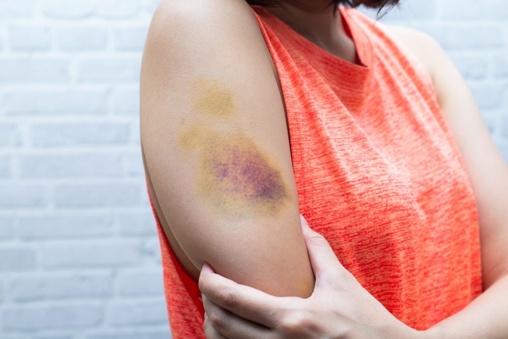
In forensic medicine, traces of foreign substances left on the body/corpse (including on its clothing and the crime scene) may be important. Examples include paint particles, lubricants, glass from a traffic accident, or gunshot residue (soot, gunpowder particles, etc.) on the body surface or clothing, which can determine the distance of the shot, the type of weapon, and identify entry and exit wounds. Biological materials such as urine, blood, sweat, semen, saliva, etc., can be traces from the perpetrator (in sexual assault, homicide) or from the victim on the perpetrator or the weapon, linking the victim to the perpetrator or the injury to the weapon. Similarly, threads and fibers from different materials, clothes, and hair – often small and invisible to the naked eye – can serve as evidence. All these traces must be identified, isolated, and collected as evidence without altering their essential characteristics – ideally without damaging their identifying features. These traces are often the most important and sometimes the only evidence in an investigation. It is crucial to find and identify them.
Special light sources have been developed, emitting light at different intensities from the visible spectrum, infrared, and ultraviolet light. Combined with various optical filters, these light sources enable the detection of different types of traces. In practice, they are most commonly used by forensic investigators at crime scenes or in laboratories as single sources of specific spectrum light for particular purposes, with each type of trace often requiring a specific source and filter. This requires good knowledge of the optical properties of different materials, good and extensive equipment, and special conditions.
In forensic medicine, particularly for biological traces or traces on corpses and living persons, it is crucial to easily and quickly detect them on-site, allowing for their collection as evidence during the inspection and autopsy process, so the work can continue, or the living patient can leave. Medical professionals do not have extensive knowledge of specific physics and do not have time to test and use multiple light sources and filters. There are two practical options:
- Use of One or Two Types of Monospectral Equipment: For the most common traces, this option limits possibilities and creates a high risk of missing important traces.
- Use of Multispectral Light Sources with Multiple Filters: Combined into one device, this option allows for quick and comprehensive examination of the body and other objects, as well as real-time evidence collection (e.g., during patient examination for sexual assault or before proceeding to autopsy after external examination of the corpse).
Various specific applications are possible depending on the circumstances, objectives, available time, and the technical and budgetary capabilities of the institution where the work is performed. Of course, specific additional training or education is required depending on the needs and the technology used.
Dr. Yanko G. Kolev, MD, PhD