Vaginal examination is a crucial part of sexual assault investigations. It is critical that the healthcare professionals performing the examination recognize normal anogenital anatomy, use the proper technique and equipment, and be able to discern abnormalities that may be connected to sexual assault from typical findings. However, it should be noted that a normal vaginal examination that does not contain any traumatic findings will not eliminate the possibility of sexual assault.
A vaginal examination is often performed in the lithotomy position. Examination in the lithotomy position can be performed in the knee-elbow position to better evaluate lesions between 5 o’clock and 7 o’clock positions, clock face localization. Once the examiner pulls the labium majus and minuses towards themselves and slightly upwards, the hymen becomes visible. The type of hymen, the measured size of the hymenal aperture, the thickness and elasticity of the hymen, congenital notches, ecchymoses, abrasions, new or previously existing lacerations, whether they reach the hymenal base or not, and their clock-face localization should all be considered.
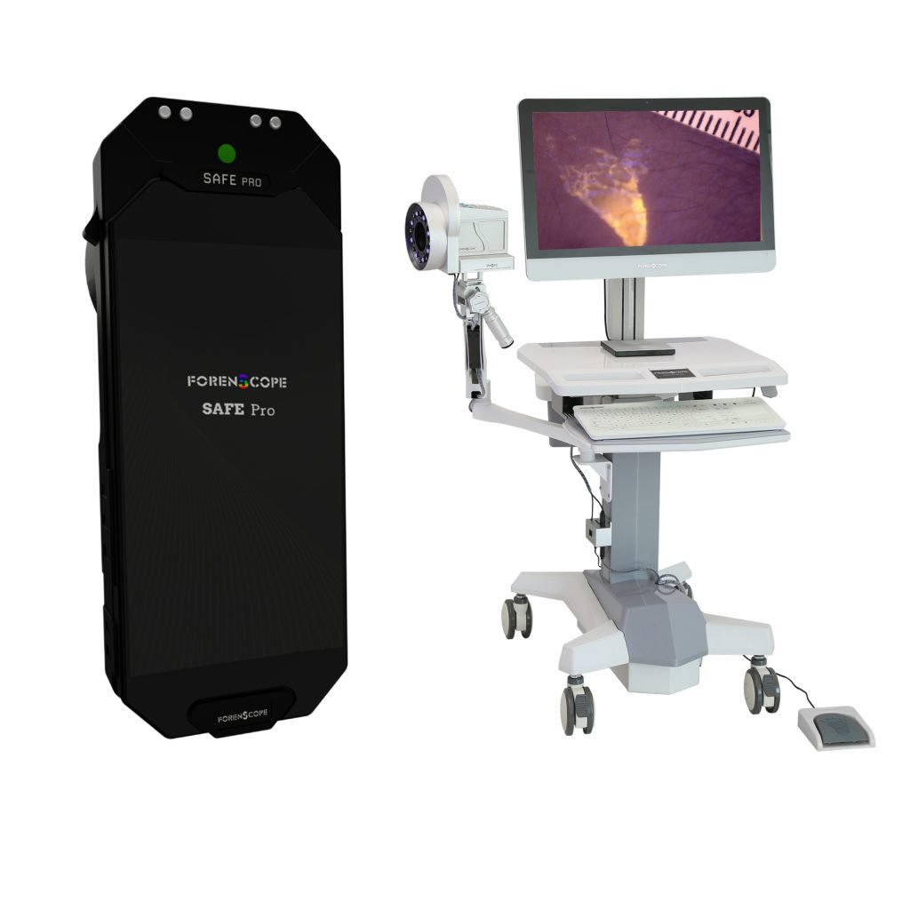
Hymen injuries are an important finding in cases of penetrative sexual assault since they are valuable as forensic evidence. Assessments performed macroscopically via the naked eye to evaluate hymen are no longer relevant, also, it might prove difficult to identify sexual assault lesions on the hymen with the naked eye. Hymen examinations assisted by SAFE (Sexual Assault Forensic Examination) Imaging Systems provide a sensitive and comprehensive assessment, enabling the identification of potential lesions that would otherwise go undetected macroscopically. SAFE (Sexual Assault Forensic Examination) Imaging Systems also make it possible to capture and store photo evidence, recording the lesions found and gathering credible proof for the legal system. Protecting the victim’s rights throughout the legal procedure is why these steps are crucial.
Lacerations often occur below the 3 o’clock and 9 o’clock positions of the clock face localization in the gynecological position. While accompanying findings such as bleeding and congestion can help distinguish acute hymen lacerations, it can be challenging to differentiate between congenital notches and preexisting hymenal tears. Congenital notches do not reach the hymenal base. It is relatively easier to differentiate between congenital notches and preexisting tears that extend to the hymenal base with a thorough examination. However, distinguishing partial tears that do not reach the hymenal base from congenital notches is quite difficult and requires expertise. The literature reports that notches are low depth, smooth-edged, and rounded, while partial tears are deep, irregular, and angular. Making this differentiation via macroscopic assessment may lead up to a large margin of error. The value of SAFE (Sexual Assault Forensic Examination) Imaging Systems lies in its ability to provide higher levels of distinction through up to apprx. 200 times increased optic and digital magnification, useful for lesions otherwise would be difficult to identify macroscopically. SAFE (Sexual Assault Forensic Examination) Imaging Systems allow for zooming in to identify notches that extend too close to the hymenal base without reaching it, leaving a thin hymenal tissue between.
Taking a swab sample, which provides biological evidence, is a crucial element of the vaginal examination. The detection of sperm in the sample is an essential finding for identifying the perpetrator. Even if the perpetrator’s sperm is present in the victim’s body, there is a good chance that no sperm will be found in samples taken during standard examinations performed with the naked eye. Sperm glows via the various light filters of SAFE (Sexual Assault Forensic Examination) Imaging Systems. This is extremely significant for indicating the area that is to be sampled. Using these samples as evidence will be highly helpful in holding perpetrators accountable for the assaults.
As a result, the use of SAFE (Sexual Assault Forensic Examination) Imaging Systems gives substantial advantages to healthcare providers in terms of preserving victim rights, providing comprehensive examinations, and contributing to the judicial process in an unbiased and authentic manner.
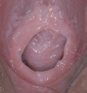
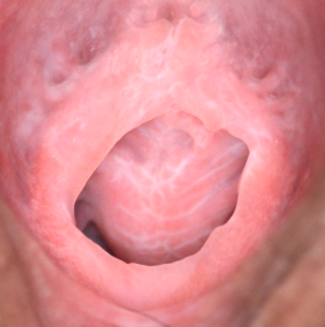
Fig 1: Intact hymen images captured by Forenscope SAFE systems with White light and Polarized light
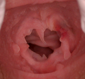
Fig 2: Incomplete laceration of hymen with ecchymosis at 2 O’clock position
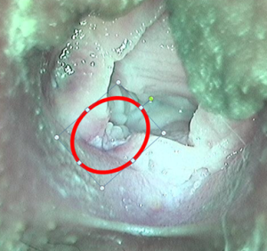
Fig 3: Inflammatory changes at the margins of hymenal laceration at healing stage captured with yellow filter of Forenscope SAFE systems
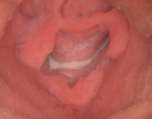
Fig 4: Old lacerations of hymen at 3 and 8 O’clock positions
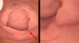
Fig 5: Complete laceration of hymen and its high magnification image captured by Forenscope SAFE systems
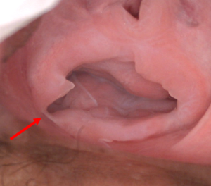
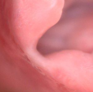
Fig 6: Complete laceration of hymen and its high magnification image captured by Forenscope SAFE systems
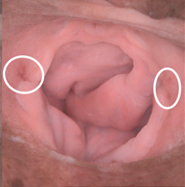
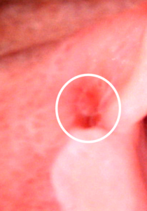
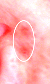
Fig 7: Hymenal ecchymosis and its high magnification image captured by Forenscope SAFE systems